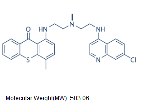
No malignancy of Mechlorethamine hydrochloride post-mitotic neurons has occurred spontaneously or been induced by carcinogens in the adult cortex. Primary heart tumors are extremely rare and so far such tumors always derive from heart cells which are not cardiomyocytes. Moreover, organisms mainly constituted by post-mitotic cells do not develop cancer as shown in adult Drosophila melanogaster, a postmitotic organism save for its germ cells, in which tumors may only arise before the larval stage, when the somatic cells are not yet TD and so preserve a proliferating potential. Indeed, adult flies subject to mutagenic radiation may die but do not develop cancer. These facts suggest that there is no set of somatic mutations able to cancel the post-mitotic state. In the interphase, nuclear DNA of metazoan cells is organized in supercoiled loops anchored to a nuclear compartment known as the nuclear matrix that is a non-soluble complex of ribonucleoproteins obtained after extracting the nucleus with high salt and treatment with DNase. The DNA is anchored to the NM by means of non-coding sequences of variable length known as matrix Gomisin-D attachment regions or MARs. Yet there is no consensus sequence for a priori identification of MARs. The DNA-NM interactions define a higher-order structure in the cell nucleus. Hepatocytes preserve a remarkable proliferating potential that can be elicited in vivo by partial hepatectomy leading to liver regeneration. We have shown that in the nucleus of primary rat hepatocytes the average DNA loop size becomes significantly reduced as a function of animal age implying the progressive increase in the number of DNA-NM interactions. This fact correlates on the one hand with a dramatic strengthening of the NM framework and the stabilization of such DNA-NM interactions and on the other hand, with reduction of  the cell proliferating potential and progression towards terminal differentiation of the hepatocytes with age. We have also compared the NHOS of aged rat hepatocytes with that of early post-mitotic rat neurons, the results indicated that a very stable NHOS is a common feature of both aged and post-mitotic cells in vivo. In the present work we compared the NHOS in rat neurons from different postnatal ages and our results show that the trend towards further stabilization of the NHOS continues in neurons beyond the fourth post-natal week, when the synapses in the cerebral cortex become indistinguishable for those in the adult rat brain and so the neurons are formally regarded as terminally differentiated. This phenomenon occurs in absence of overt changes in the post-mitotic state and transcriptional activity of neurons, suggesting that the continued stabilization of the NHOS as a function of time is basically determined by thermodynamic and structural constraints. We discuss how the resulting highly stable NHOS of neurons may be the non-genetic basis of their permanent and irreversible post-mitotic state. The rat cerebral cortex contains both neurons and glial cells. The classical method by Thompson designed for the isolation of neuronal nuclei separates two nuclear populations: N1 mostly constituted by neuronal nuclei according to old morphological criteria and N2 relatively enriched for glial-cell nuclei on morphological grounds. NeuN/Fox-3 is a protein specific of neuronal nuclei and so labelling with anti-NeuN mAb is the current standard for reliable identification of neurons and neuronal nuclei. In newborn rats the glial cells constitute a small percentage of the brain cells hence there is no point in separating N1 and N2 populations.
Primary tumors arise from the glial cells that preserve a proliferating potential or from precursor cells
Leave a reply